Fast Volumetric Imaging of Seizures with Adaptive Optics Light Sheet Microscopy
AI Summary
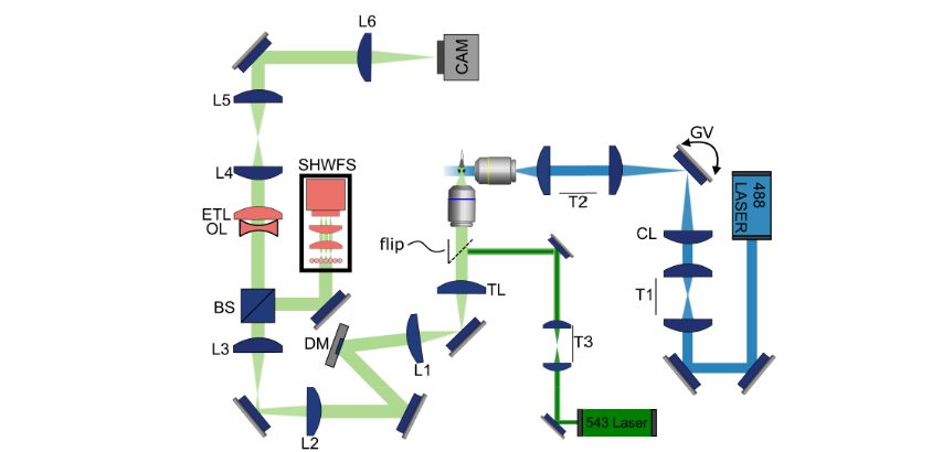
Seizure in zebrafish larva imaged using AO setup. (Credit: Bingxi Liu et al ., Biomedical Optics Express, 2026) Key to understanding something like epilepsy is to be able to record highly transient events in biological tissues. Generally this is done using light sheet microscopy, which provides effectively a 2D ‘slice’ of the tissue in question, but to observe a brief event in a larger biological system you need to be able to rapidly change the layer and focus between the virtual layers.
Discussion
Sign in to join the discussion.
Loading comments…